
Cerebrospinal fluid circulation
Cerebrospinal fluid is produced by the choroid plexuses, which are located in the lateral ventricles (along the line of the choroid fissure) and in the roof of the third and fourth ventricles. The vascular source of the choroid plexus differs between the lateral and third ventricles, and the fourth ventricle. The lateral and third ventricles are supplied anteriorly by the internal carotid and choroidal branches of the posterior cerebral artery, whereas the fourth ventricle, being much lower in position, is supplied by the inferior cerebellar arteries. The CSF is not passively filtered into the ventricles but actively secreted by the choroid plexus epithelium, which shares many characteristics with secretory and transport cells. The epithelial lining is a complex system: the capillaries are tortuous, presenting interdigitations, while the apical, secretory borders contain microvilli; the vessels are fenestrated underneath the epithelial lining, with tight junctions in the apical border of the epithelia, allowing only small molecules and fluid through. In this way, the choroid plexus is still able to maintain a Blood-CSF barrier, while still supplying fluid to cushion and nurture the brain.
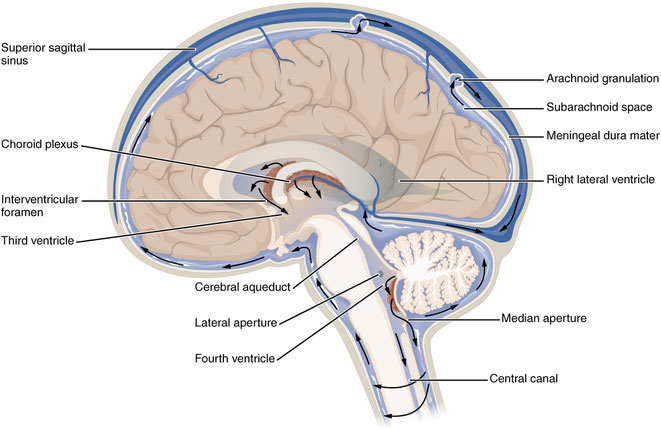
Cerebrospinal fluid is constantly produced at a secretion rate of 0.35-0.40 mL.min-1, and replaced with an average volume of 150 mL in the ventricular system/subarachnoid space. Of this volume, about 125 mL is intracranial and 25 mL of this volume lies within the ventricles. The circulating CSF flows from the lateral ventricles through the third and fourth ventricles and into the subarachnoid space. From the fourth ventricle, the CSF either continues to the central canal of the spinal cord or leaves for the subarachnoid space by passing through the medial aperture (foramen of Magendie) and paired lateral apertures (foramina of Luschka) in the roof and lateral recesses of the fourth ventricle, respectively. The level at which the CSF enters the subarachnoid space is called the cerebellomedullary cistern. The fluid then flows through the subarachnoid space surrounding the brain and spinal cord. It finally leaves the subarachnoid space and CNS by entering the venous sinuses through the arachnoid villi. The arachnoid villi provide a valvular mechanism for flow of CSF into the bloodstream without permitting backflow of blood into the CSF.