Cerebrum, cerebellum, and brain stem
Cerebrum
The cerebrum is the largest part of the brain, located superiorly and anteriorly in relation to the brainstem. It consists of two cerebral hemispheres (left and right), separated by the falx cerebri of the dura mater. Embryologically, the cerebrum is derived from the telencephalon.
The cerebrum is comprised of two different types of tissue – grey matter and white matter:
- Grey matter forms the surface of each cerebral hemisphere (known as the cerebral cortex), and is associated with processing and cognition.
- White matter forms the bulk of the deeper parts of the brain. It consists of glial cells and myelinated axons that connect the various grey matter areas.
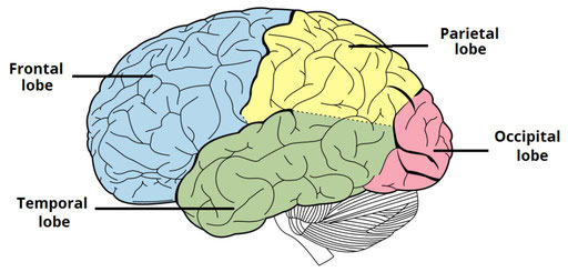
The cerebral cortex is classified into four lobes, according to the name of the corresponding cranial bone that approximately overlies each part. Each lobe contains various cortical association areas – where information from different modalities are collated for processing. Together, these areas function to give us a meaningful perceptual interpretation and experience of our surrounding environment.

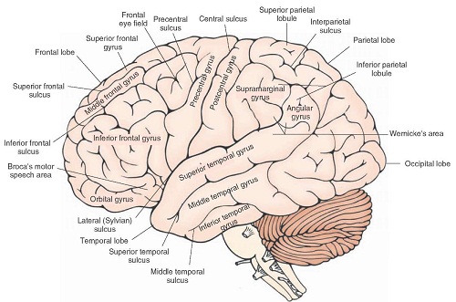
Externally, the cerebrum has a highly convoluted appearance, consisting of sulci (grooves or depressions) and gyri (ridges or elevations). It is divided into two anatomically symmetrical hemispheres by the longitudinal fissure – a major sulcus that runs in the median sagittal plane. The falx cerebri (a fold of dura mater) descends vertically to fill this fissure. The two cerebral hemispheres are connected by a white matter structure, called the corpus callosum.
The main sulci are:
- Central sulcus – groove separating the frontal and parietal lobes.
- Lateral sulcus – groove separating the frontal and parietal lobes from the temporal lobe.
- Lunate sulcus – groove located in the occipital cortex.
The main gyri are:
- Precentral gyrus – ridge directly anterior to central sulcus, location of primary motor cortex.
- Postcentral gyrus – ridge directly posterior to central sulcus, location of primary somatosensory cortex.
Superior temporal gyrus – ridge located inferior to lateral sulcus, responsible for the reception and processing of sound.
Cerebellum
The cerebellum is a structure of the central nervous system situated in the posterior cranial fossa. It lies posterior to the pons and medulla, separated from them by the fourth ventricle. The cerebellum plays an important role in motor function, especially in coordination, precision, timing of movement, and motor learning.
The cerebellum is spherical in shape, and consists of two large hemispheres united in the middle by the vermis. It is divided by numerous transverse fissures into lobes and lobules. The three lobes of the cerebellum are the anterior, posterior, and flocculonodular lobe, which consists of a flocculus and a nodule. The deepest fissure of the cerebellum is termed the primary fissure. It marks the boundary between the anterior and posterior lobes. Another prominent fissure is the horizontal fissure, which separates the cerebellum into superior and inferior surfaces.


Click to enlarge image. Click to enlarge image.
The cerebellum is comprised of a gray cerebellar cortex, a medullary core of white matter, and four pairs of intrinsic nuclei. The cerebellar cortex consists of numerous narrow leaf-like laminae known as the cerebellar folia. These folia are parallel with each other, and are transversely oriented. Moreover, each lamina includes several secondary and tertiary folia as well. The medullary core of white matter has a complex branching system, and together with the tree-like appearance of the laminae and folia, it has been given the descriptive term ‘arbor vitae’, the tree of life.
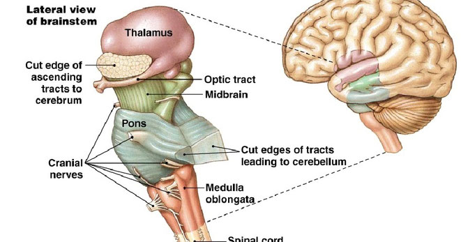
Brain stem
The brainstem is the structure situated at the base of the brain, connecting the deep structures of the cerebral hemispheres and the cervical portion of the spinal cord. It is associated with various vital functions of the body. The brainstem houses the majority of the cranial nerve nuclei, except those involved with olfaction and vision. These nuclei provide motor and sensory functions to the structures of the cranium. The brainstem also consists of important nuclei associated with parasympathetic and sympathetic functions. Moreover, efferent and afferent pathways between the cerebrum and cerebellum course through the brainstem, where they mostly decussate or cross each other. The brainstem is formed of three parts: the medulla oblongata, the pons and the midbrain.

The medulla oblongata is the lowest portion of the brainstem. It is continuous with the spinal cord from below, and the pons from above. The medulla is responsible for various autonomic functions and contains cardiac, respiratory, reflex, and vasomotor centers.
The pons is the middle portion of the brainstem, lying between the medulla and the midbrain. It is mainly involved with sleep, respiration, swallowing, hearing, bladder control, equilibrium, and taste, as well as various other motor functions.
The midbrain is the most upper portion of the brainstem. It lies between the pons from below, and the thalamus from above. It plays an important role in motor eye movement, visual and auditory processing, in addition to alertness and temperature regulation. The cerebellum is connected to the brainstem by three pairs of cerebellar peduncles: the superior peduncle with the midbrain, the middle peduncle with the pons, and the inferior peduncle with medulla oblongata.