
Vascular anatomy
The brain is supplied by branches of the internal carotid artery anteriorly and by branches of the vertebral artery posteriorly.
The aortic arch gives off three great vessels: the brachiocephalic artery, the left common carotid artery and the left subclavian artery. The brachiocephalic artery subsequently divides into the right common carotid artery and right subclavian artery.

Within the mid portion of the neck, the common carotid artery splits into the internal carotid artery (which goes directly to the brain) and the external carotid artery (which gives off a number of branches that supply the neck and face).


The internal carotid artery enters the skull base through the carotid canal within the petrous portion of the temporal bone and ascends within the cavernous sinus. Once it exits the cavernous sinus it courses intracranially over the anterior clinoid process and terminates in a T junction giving rise to the anterior cerebral artery medially and the middle cerebral artery laterally.
The vertebral artery arises from the subclavian artery. The paired vessels ascend through the neck within the foramen transversarium of the cervical spine. This relationship is important to understand as patients with cervical spine trauma may have fractures that involve the foramen, putting them at risk for vertebral artery injury. The vertebral artery is divided into four parts and its origin (V0-V4) as shown in the figure below.

The Circle of Willis
The Circle of Willis is an arterial polygon formed as the internal carotid and vertebral systems anastomose around the optic chiasm and infundibulum of the pituitary stalk in the suprasellar cistern. This communicating pathway allows equalization of blood-flow between the two sides of the brain, and permits anastomotic circulation, should a part of the circulation be occluded.


Click image to enlarge. Click image to enlarge.
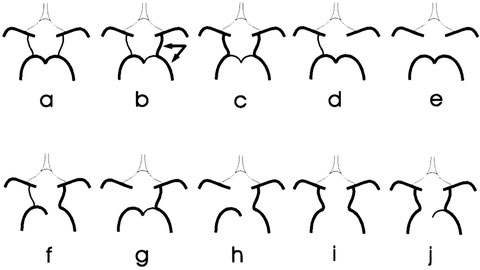
A complete circle of Willis (in which no component is absent or hypoplastic) is only seen in 20-25% of individuals. Posterior circulation anomalies are more common than anterior circulation variants and are seen in nearly 50% of anatomical specimens.
Common variants:
- hypoplasia of one or both PCOM ~30% (range 25-34%)
- hypoplastic/absent A1 segment of ACA ~15% (range 10-15%)
- absent or fenestrated ACOM ~12.5% (range 10-15%)
- origin of PCA from the ICA with absent/hypoplastic P1 segment (fetal PCOM) ~20% (range 17-25%)
- infundibular dilatation of the PCOM origin ~10% (range 5-15%)
Congenital absence of one or both ICAs may occur but is rare. If one ICA is absent, intrasellar intercarotid communicating arteries are common and there is a high incidence of associated aneurysms.
For more information on Circle of Willis variations see:
https://www.ncbi.nlm.nih.gov/pmc/articles/PMC3879841/pdf/jcdr-7-2423.pdf
Middle cerebral arteries
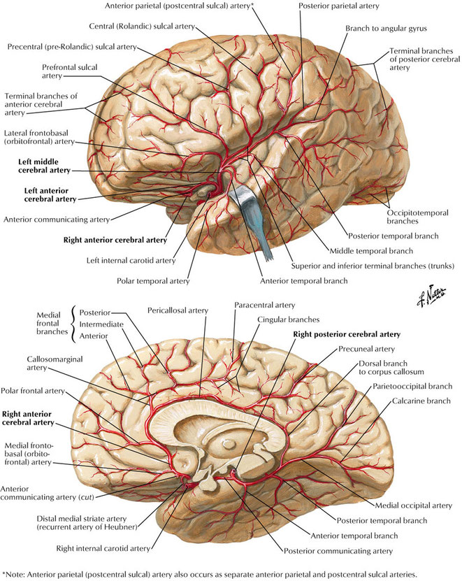
The middle cerebral artery (MCA) is one of the three major paired arteries that supply blood to the brain. The MCA arises from the internal carotid artery (ICA) as the larger of the two main terminal branches (the other being the anterior cerebral artery), coursing laterally into the lateral sulcus where it branches and provides many branches that supply the cerebral cortex.
Segments
The MCA is divided into four segments:
- M1: from the origin to bifurcation/trifurcation (the limen insulae); also known as the horizontal or sphenoidal segment.
- M2: also known as the insular segment, from the bifurcation/trifurcation to the circular sulcus of the insula, where it makes a hairpin bend to continue as M3.
- M3: opercular branches (those within the Sylvian fissure); also known as the opercular segment.
- M4: branches emerging from the Sylvian fissure onto the convex surface of the hemisphere; also known as the cortical segment.
Supply
The middle cerebral arteries supply the majority of the lateral surface of the hemisphere, except the superior portion of the parietal lobe (via the ACA) and the inferior portion of the temporal lobeand occipital lobe (via the PCA). In addition, they supply part of the internal capsule and basal ganglia.
In its territory lie the motor and sensory areas excluding leg and perineum and auditory and speech areas.
Anterior cerebral arteries
The anterior cerebral artery along with the middle cerebral artery forms at the termination of the internal carotid artery. It is the smaller of the two, and arches anteromedially to pass anterior to the genu of the corpus callosum, dividing as it does so into its two major branches; pericallosal and callosomarginal arteries. It supplies the medial aspect of the cerebral hemispheres back to the parietal lobe.
Segments
The ACA is divided into three segments:
- A1 (horizontal): origin from the ICA to the anterior communicating artery (ACOM), ~14 mm in length
- A2 (vertical): from ACOM to the origin of the callosomarginal artery
- A3 (callosal): distal to the origin of the callosomarginal artery
Posterior cerebral arteries
The posterior cerebral arteries (PCA) are the terminal branches of the basilar artery. It curls back around the cerebral peduncle and pass above the tentorium, to supply the occipital lobes and posteromedial temporal lobes.
Segments
- P1 (mesencephalic): from it origin at the termination of the basilar artery to posterior communicating artery (PCOM), within interpeduncular cistern. Perforating branches arise from P1.
- P2 (ambient): from the PCOM around the cerebral peduncle above occulomotor, divided into P2A (anterior) and P2P (posterior) sub-segments. P2A is within crural cistern which then bridges to the P2P segment in ambient cistern (thus ambient segment).
- P3: quadrigeminal segment (within the quadrigeminal cistern) extends posteromedially from level of quadrigeminal plate.
- P4: cortical segment (e.g. calcarine artery, within the calcarine fissure) arise from distal PCA at or just before reaching calcarine fissure.
Supply
Penetrating branches: Midbrain, thalami, posterior limb of internal capsule, optic tract
Ventricular/choroidal branches: Choroid plexus of third/lateral ventricles, parts of thalami, posterior commissure, cerebral peduncles
Splenial branches: Posterior body and splenium of corpus callosum
Cortical branches: Posterior 1/3 of medial hemisphere surface; most of inferior temporal lobe, most of occipital lobe (including visual cortex).
Branches
- P1:
Posterior thalamoperforating arteries: pass posterosuperiorly in interpeduncular fossa and enter undersurface of midbrain.
- P2:
Thalamogeniculate arteries: pass posteromedially into midbrain
Peduncular perforating arteries: pass directly into cerebral peduncles
Medial/lateral posterior choroidal branches: pass around medial geniculate body to reach the posterior part of the inferior horn of the lateral ventricle to supply the choroid plexus.
- PCOM:
anterior thalamoperforator.
- P4:
Two terminal trunks:
Lateral: posterior temporal arteries supplies the posterior part of the temporal lobe.
Medial: medial occipital artery, divide into calcarine artery and parieto-occipital artery, splenial artery.