
Vertebral column
Vertebral column
The vertebral column (spinal column) is a vertical series of approximately 33 small bones (known as vertebrae), which are separated by intervertebral discs. It can be separated into five different regions, with each region characterised by a different vertebral structure. All vertebrae share a basic common structure. They each consist of a vertebral body, situated anteriorly, and a posterior vertebral arch.
The vertebral body is the anterior part of the vertebrae. It is the weight-bearing component, and its size increases as the vertebral column descends (having to support increasing amounts of weight).
The superior and inferior aspects of the vertebral body are lined with hyaline cartilage. Adjacent vertebral bodies are separated by a fibrocartilginous intervertebral disc.
The vertebral arch refers to the lateral and posterior parts of the vertebrae.
With the vertebral body, the vertebral arch forms an enclosed hole, called a vertebral foramen. The foramina of the all vertebrae line up to form the vertebral canal, which encloses the spinal cord.
The vertebral arches have a number of bony prominences, which act as attachment sites for muscles and ligaments:
- Pedicles: There are two of these, one left and one right. They point posteriorly, meeting the flatter laminae.
- Lamina: The bone between the transverse and spinal processes.
- Transverse processes: These extend laterally and posteriorly away from the pedicles. In the thoracic vertebrae, the transverse processes articulate with the ribs.
- Articular processes: At the junction of the lamina and the pedicles, superior and inferior processes arise. These articulate with the articular processes of the vertebrae above and below.
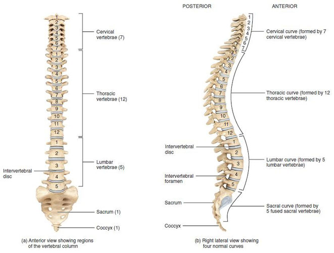
Cervical
There are seven cervical vertebrae in the human body. They have three main distinguishing features:
- The spinous process bifurcates into two parts, and so is known as a bifid spinous process.
- There are two transverse foramina, one in each transverse process. These conduct the vertebral arteries.
- The vertebral foramen is triangular in shape
There are some cervical vertebrae that are unique. C1 and C2 (called the atlas and axis respectively), are specialised to allow for the movement of the head.
The C7 vertebrae has a much longer spinous process, which does not bifurcate.

Thoracic
The twelve thoracic vertebrae are medium-sized, and increase in size as they move down the back. Their main function is to articulate with ribs, producing the bony thorax.
Each thoracic vertebrae has two ‘demi facets‘ on each side of its vertebral body. These articulate with the head of the respective rib, and the rib inferior to it. On the transverse processes of the thoracic vertebrae there is a costal facet for articulation with its respective rib.
The spinous processes are slanted inferiorly and anteriorly. This offers increased protection to the spinal cord, preventing an object like a knife entering the spinal canal through the intervertebral discs.
In contrast to the cervical vertebrae, the vertebral foramen is circular.

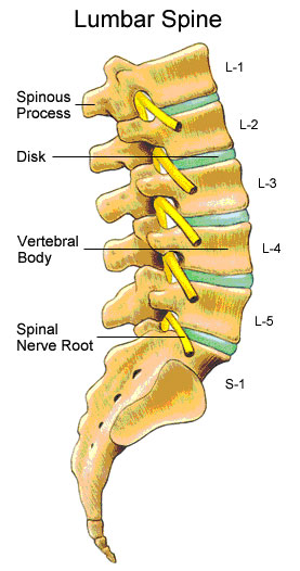
Lumbar
These are the largest of the vertebrae, of which there are five (or six). They act to support the weight of the upper body, and have various specialisations to enable them do this.
Lumbar vertebrae have very large vertebral bodies, which are kidney-shaped. They lack the characteristic features of other vertebrae, with no transverse foramina, costal facets, or bifid spinous processes.
Like the cervical vertebral, they have a triangular shaped vertebral foramen.
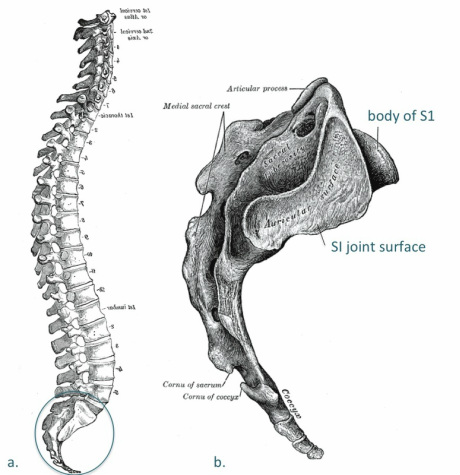
Sacrum and Coccyx
The sacrum is a collection of five fused vertebrae. It is described as an upside down triangle, with the apex pointing inferiorly. On the lateral walls of the sacrum are facets, for articulation with the pelvis at the sacro-iliac joints.
The coccyx is a small bone, which articulates with the apex of the sacrum. It is recognised by its lack of vertebral arches. Due to the lack of vertebral arches, there is no vertebral canal, and so the coccyx does not transmit the spinal cord
Joints
Each vertebra has five articulations. The vertebral bodies indirectly articulate with each other, and the articular processes also form joints. The vertebral body joints are cartilaginous joints, designed for weight-bearing. The articular surfaces are covered by hyaline cartilage, and are connected by a fibrocartilage intervertebral disc. There are two ligaments that strengthen these joints; the anterior and posterior longitudinal ligaments. The anterior longitudinal ligament is thick and prevents hyperextension of the vertebral column. The posterior longitudinal ligament is weaker and prevents hyperflexion.
The joints between the articular facets are called facet joints. These allow for some gliding motions between the vertebrae. They are strengthened by various ligaments:
- Ligamentum Flavum: extends from lamina to lamina.
- Interspinous and Supraspinous ligaments: These join the spinous processes together. The interspinous ligaments attach between processes, and the supraspinous ligaments attach to the tips.
- Intertransverse ligaments: extends between transverse processes.