
Vestibulocochlear nerve
The vestibular and cochlear portions of the vestibulocochlear nerve are functionally discrete, and so originate from different nuclei in the brain:
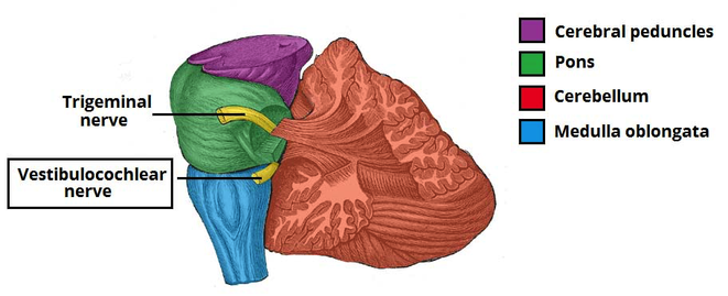
- Vestibular component – arises from the vestibular nuclei complex in the pons and medulla.
- Cochlear component – arises from the ventral and dorsal cochlear nuclei, situated in the inferior cerebellar peduncle.
Both sets of fibres combine in the pons to form the vestibulocochlear nerve. The nerve emerges from the brain at the cerebellopontine angle and exits the cranium via the internal acoustic meatus of the temporal bone.
Within the distal aspect of the internal acoustic meatus, the vestibulocochlear nerve splits, forming the vestibular nerve and the cochlear nerve. The vestibular nerve innervates the vestibular system of the inner ear, which is responsible for detecting balance. The cochlear nerve travels to cochlea of the inner ear, forming the spiral ganglia which serve the sense of hearing.
The vestibulocochlear nerve is unusual in that it primarily consists of bipolar neurones. It is responsible for the special senses of hearing (via the cochlear nerve), and balance (via the vestibular nerve).
Hearing
The cochlea detects the magnitude and frequency of sound waves. The inner hair cells of the organ of Corti activate ion channels in response to vibrations of the basilar membrane. Action potentials travel from the spiral ganglia, which house the cell bodies of neurones of the cochlear nerve.
The magnitude of the sound determines how much the membrane vibrates and thereby how often action potentials are triggered. Louder sounds cause the basilar membrane to vibrate more, resulting in action potentials being transmitted from the spiral ganglia more often, and vice versa. The frequency of the sound is coded by the position of the activated inner hair cells along the basilar membrane.
Equilibrium (Balance)
The vestibular apparatus senses changes in the position of the head in relation to gravity. The vestibular hair cells are located in the otolith organs (the utricule and saccule), where they detect linear movements of the head, as well as in the three semicircular canals, where they detect rotational movements of the head. The cell bodies of the vestibular nerve are located in the vestibular ganglion which is housed in the outer part of the internal acoustic meatus.
Information about the position of the head is used to coordinate balance and the vestibulo-ocular reflex. The vestibulo-ocular reflex (also called the oculocephalic reflex) allows images on the retina to be stabilised when the head is turning by moving the eyes in the opposite direction. It can be demonstrated by holding one finger still at a comfortable distance in front of you and twisting your head from side to side whilst staying focused on the finger.

Clinical relevance
A lesion on the vestibular nerve would result in nystagmus (rhythmic oscillations of the pupil), unsteady gait, nausea and vertigo. A lesion on the auditory nerve would result in hearing loss and/or tinnitus.
- One of the most common brain tumors, a vestibular schwannoma or acoustic neuroma, occurs on the vestibular portion of cranial nerve VIII and is marked by the many of the above mentioned signs and symptoms. Many patients complain on tinnitus in one ear with fullness in that ear. Although acoustic neuromas are typically slow growing and benign, in rare cases they may grow large enough to compress the brainstem and threaten the patient’s life.
- The vestibulocochlear nerve can be damaged due to a basilar skull fracture, usually resulting from major head trauma. The nerve may be damaged in the internal acoustic meatus resulting in vestibular and cochlear symptoms.
- Vestibular neuritis refers to inflammation of the vestibular branch of the vestibulocochlear nerve. The aetiology of this condition is not fully understood, but some cases are thought to be due to reactivation of the herpes simplex virus. It presents with symptoms of vestibular nerve damage. The condition is usually self-resolving. Treatment is symptomatic, usually in the form of anti-emetics or vestibular suppressants.
- Labyrinthitis refers to inflammation of the membranous labyrinth, resulting in damage to the vestibular and cochlear branches of the vestibulocochlear nerve.
The symptoms are similar to vestibular neuritis, but also include indicators of cochlear nerve damage like sensoneurinal hearing loss and tinnitus.