
Optic nerve
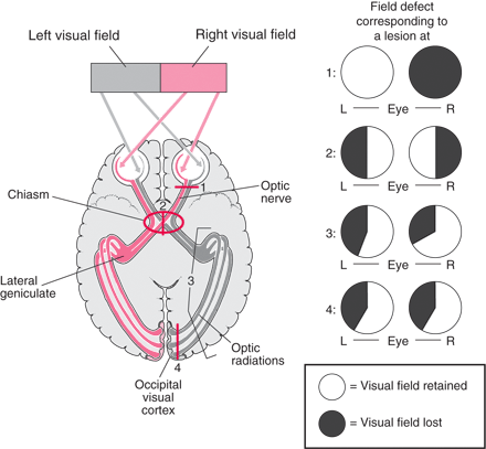
Nerve signals travel along the optic nerve from each eye. The two optic nerves meet at the optic chiasm. There, the optic nerve from each eye divides, and half of the nerve fibers from each side cross to the other side. Because of this arrangement, the right side of the brain receives information from the left visual field of both eyes, and the left side of the brain receives information from the right visual field of both eyes. Damage to an eye or the visual pathway causes different types of vision loss depending on where the damage occurs.
Aetiology of optic nerve disorders
- Congenital anomalies
- Glaucoma
- Inflammatory (optic neuritis)
- Ischaemic
- Raised intracranial pressure (papilloedema)
- Compression
- Nutritional and toxic optic neuropathy
- Trauma
- Inherited optic neuropathy
- Infiltrative (neoplastic or granulomatous)
- Optic atrophy secondary to retinal disease
Rarely can the aetiology of optic neuropathy be made on the basis of a single clinical finding. There is considerable overlap of clinical features between even the relatively common optic neuropathies, from which arises the potential value of neural networks. However, in experienced hands clinical diagnosis is reliable and sufficient to avoid or minimise further investigation. Recognition of retinal disease or nonorganic visual loss masquerading as optic neuropathy is particularly important.
Temporal course
Clarification of the temporal course of visual loss is critical. Ischaemic optic neuropathy generally has a sudden onset, although in some cases there is progressive visual loss over a period of 2 weeks, with moderate recovery at best thereafter. Inflammatory optic neuropathy tends to have a subacute onset over a number of days, frequently accompanied by periocular pain exacerbated by eye movements. The almost complete recovery of vision over the next few weeks is sufficiently characteristic of the vast majority of cases of demyelinating optic neuropathy to allow a clinical diagnosis, without the need for confirmatory investigation as long as there are no atypical features. Compressive optic neuropathy usually causes gradually progressive visual loss, although particularly vascular lesions may cause abrupt visual decline occasionally followed by some recovery. Nutritional or toxic optic neuropathy results in subacute or chronic symmetrical bilateral visual loss. Visual loss due to congenital anomalies rarely changes, except in optic disc pits complicated by serous macular detachment.
Glaucomatous optic neuropathy is not symptomatic until late in the disease course such that it is usually identified incidentally or by screening. In Leber's hereditary optic neuropathy, visual loss most commonly develops sequentially with 8 weeks delay between involvement of the two eyes and progression of visual loss over 4–6 weeks in each eye. Dominant optic atrophy causes very gradually progressive or nonprogressive visual loss.
Negative and positive visual phenomena
In general, optic nerve disease produces negative and retinal disease positive visual phenomena, but photopsiae do occur in optic neuropathy. Micropsia, macropsia, and central visual distortion strongly suggest macular dysfunction. Night blindness indicates rod dysfunction, although many patients with optic nerve dysfunction complain of particularly poor vision in dim light. Deterioration of vision with increased body temperature either from external heating or induced by exercise (Uhthoff's phenomenon) most commonly occurs in demyelinating optic neuropathy, especially in primary progressive multiple sclerosis, but it may occur with many types of optic neuropathy. Gaze-evoked amaurosis characteristically occurs with optic nerve sheath meningioma but may be produced by other lesions deforming the optic nerve, including primarily extraorbital disease.
Ischaemic optic neuropathy in a patient aged over 50 years necessitates exclusion of giant cell arteritis. Jaw claudication is almost pathognomonic. Recent onset severe headache, scalp tenderness, polymyalgia rheumatica, and general malaise are important features. Confusion and angina are serious features indicating cerebral and coronary insufficiency. It also needs to be borne in mind that nonarteritic anterior ischaemic optic neuropathy rarely occurs in optic discs with large cups.
Clinical assessment of optic nerve disorders https://www.nature.com/eye/journal/v18/n11/pdf/6701575a.pdf