
Meninges
The meninges refer to the membranous coverings of the brain and spinal cord. There are three layers of meninges, known as the dura mater, arachnoid mater and pia mater.
These coverings have two major functions:
- Provide a supportive framework for the cerebral and cranial vasculature.
- Acting with cerebrospinal fluid to protect the CNS from mechanical damage.
The meninges are often involved cerebral pathology, as a common site of infection (meningitis), and intracranial bleeds

The dura mater is the outermost layer of the meninges, lying directly underneath the bones of the skull and vertebral column. It is thick, tough and inextensible. Within the cranial cavity, the dura contains two connective tissue sheets:
- Endosteal layer – Lines the inner surface of the bones of the cranium.
- Meningeal layer – Lines the endosteal layer inside the cranial cavity. It is the only layer present in the vertebral column.
Between these two layers, the dural venous sinuses are located. They are responsible for the venous vasculature of the cranium, draining into the internal jugular veins. In some areas within the skull, the meningeal layer of the dura mater folds inwards as dural reflections. They partition the brain, and divide the cranial cavity into several compartments. For example, the tentorium cerebelli divides the cranial cavity into supratentorial and infratentorial compartments.
The dura mater receives its own vasculature; primarily from the middle meningeal artery and vein. It is innervated by the trigeminal nerve (V1, V2 and V3).
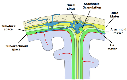
The arachnoid mater is the middle layer of the meninges, lying directly underneath the dura mater. It consists of layers of connective tissue, is avascular, and does not receive any innervation.
Underneath the arachnoid is a space known as the sub-arachnoid space. It contains cerebrospinal fluid, which acts to cushion the brain. Small projections of arachnoid mater into the dura (known as arachnoid granulations) allow CSF to re-enter the circulation via the dural venous sinuses.
The pia mater is located underneath the sub-arachnoid space. It is very thin, and tightly adhered to the surface of the brain and spinal cord. It is the only covering to follow the contours of the brain (the gyri and fissures).
Like the dura mater, it is highly vascularised, with blood vessels perforating through the membrane to supply the underlying neural tissue.