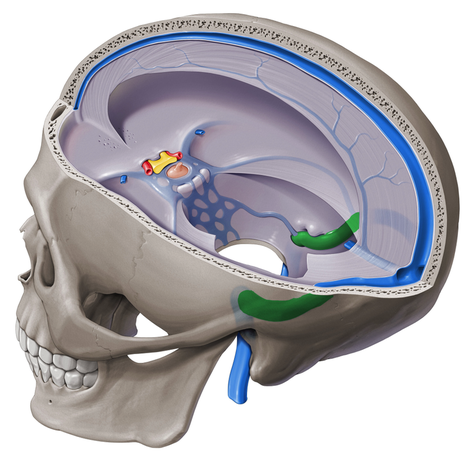
Dural venous sinuses
There are seven paired (transverse, cavernous, greater & lesser petrosal, sphenoparietal, sigmoid and basilar) and five unpaired (superior & inferior sagittal, straight, occipital and intercavernous) dural sinuses.
There are two sagittal sinuses that occupy the longitudinal cerebral fissure (midline between the cerebral hemispheres). The superior sagittal sinus is the more superficial of the two sinuses.
It resides in the base of the falx cerebri and commences at the foramen caecumof the frontal bone. It follows the contour of the calvaria (along its midline) to its termination at the internal occipital protuberance. The superior sagittal sinus receives tributaries form several superior cerebral veins that run deep to the arachnoid mater on both hemispheres. The veins pierce the arachnoid and dura as they approach the sagittal sinus, where they will drain their contents.

Deep to the superior sagittal sinus lays an inferior sagittal sinus in the free border of the falx cerebri, just dorsal to the corpus callosum. The sinus is also concaved as, but smaller than, its superior counterpart. It extends from the region just above the cribriform plate of the ethmoid bone and continues to the anterior midline of the tentorium cerebelli. Along its journey, it receives tributaries that drain from the medial part of the cerebral hemispheres. At the tentorium cerebelli, it drains into the straight sinus.
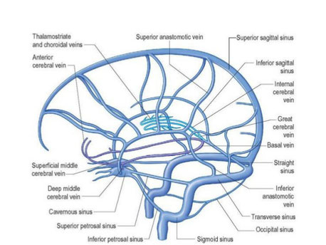
Each anterior cerebral vein leaves the longitudinal cerebral fissure inferiorly and crosses the optic chiasm ventrally to join the respective deep middle cerebral(which coursed medially through the lateral sulcus of Sylvius). Together, these vessels form the basal vein of Rosenthal that also receives tributaries from the anterior pontomesencephalic (draining from the anterior pons), lateral mesencephalic veins (from the superior pons) and branches from the superior colliculi.
Click image to enlarge.
The paired internal cerebral veins (formed from the thalamostriate and choroidal veins) merge with the basal vein of Rosenthal, the posterior mesencephalic veins and the dorsal vein of corpus callosum to form the great cerebral vein of Galenat the end of the splenium of corpus callosum. The great cerebral vein of Galen then drains to the straight sinus. The straight sinus is a midline structure that lies within the posterior end of the falx cerebri and the middle of the tentorium cerebelli. It also terminates at the level of the internal occipital protuberance.
The sphenoparietal sinus courses along the free border of the lesser wing of the sphenoid bone. It follows the groove of the bone and drains into the cavernous sinus after receiving a tributary on either side from the superficial middle cerebral vein. The superficial middle cerebral vein communicates with the superior cerebral veins via the superior anastomotic vein of Trolard. It therefore aids in draining blood from the cerebral hemispheres as well as the optic part of the frontal lobe.

In addition to receiving tributaries from the sphenoparietal sinus, the cavernous sinus also receives tributaries from the superior and inferior ophthalmic veins, and efferent hypophyseal veins. This pair of sinuses, also called the parasellar sinuses, is located on either side of the sella turcica of the sphenoid bone. Their cavities have a spongy appearance that is formed by the numerous vessels passing through it.In addition to acting as a conduit for blood, the cavernous sinus also facilitates the passage of the:
- CN III (oculomotor)
- CN IV (trochlear)
- CN V1 (ophthalmic division of trigeminal)
- CN V2 (maxillary division of trigeminal)
- CN VI (abducent)
- The cavernous part of the internal carotid artery
The cavernous sinus is the only one of the paired dural sinuses that communicates with each other. These sinuses have two intercavernous branches arching over the diaphragma sellae of the pituitary gland; one anterior and the other posterior to the infundibulum. They drain posteriorly by the petrosal sinuses.
There are four petrosal sinuses leaving the middle cranial fossa. Two superior petrosal sinuses (one on either side) run along the petrous part of the temporal bone in the base of the tentorium cerebelli. It crosses over the trunk of the trigeminal nerve before it enters Meckel’s cave. It terminates in the proximal part of the sigmoid sinus.
Similarly, an inferior petrosal sinus leave the posterior extent of each cavernous sinus to travel inferolaterally towards the distal part of the sigmoid sinus inferior to the fibers of CN VII (facial) and CN VIII (vestibulocochlear).

The left and right transverse sinuses travel in the base of the tentorium cerebelli, along the occipital bone. It communicates with the straight sinus, superior sagittal sinus and the occipital sinus at a point called the confluence of sinuses; at the level of the internal occipital protuberance. The sinus also receives venous contributions from the inferior cerebral vein and the inferior anastomotic vein of Labbé, which communicates with the vein of Trolard and the superficial middle cerebral vein.
Finally, the sigmoid sinuses are a paired, bilateral, s-shaped set of sinuses that course along the floor of the posterior cranial fossa. They are the terminal parts of the dural venous sinuses that continue from the transverse sinuses at the level of the tentorium cerebelli. Just after the sigmoid sinus receives the inferior petrosal sinus, it forms a jugular bulb that enters the jugular foramen with CN IX (glossopharyngeal), CN X (vagus) and CN XI (accessory) as the internal jugular vein.