Arachnoid cysts
Arachnoid cysts are benign, usually asymptomatic cysts occurring in the central nervous system. They are commonly located intracranially, but can be found along the spinal cord as well. They are usually located in the subarachnoid space and contain CSF.
On imaging, they are characterised as well circumscribed cysts, with an imperceptible wall, displacing adjacent structures, and following the CSF pattern. They can also have a remodelling effect on the adjacent bone.
Arachnoid cysts are thought to arise due to the congenital splitting of the arachnoid layer with accumulation of CSF within this potential space. The cyst wall is comprised of flattened arachnoid cells forming a thin translucent membrane. There is no solid component and no epithelial lining.
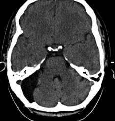
CT features
Arachnoid cysts are extremely well circumscribed, with an imperceptible wall, and displace adjacent structures. When large, and over time, they can exert a remodelling effect on the bone.
CT cisternography (introduction of contrast into the subarachnoid space) demonstrates communication of the cyst with the subarachnoid space. As this communication is slow, the cyst often fills later, and contrast may be seen to pool with it, outlining its dependent portion.