Colloid cysts
Colloid cysts are benign, congenital epithelium-lined cysts that most commonly occur in the third ventricle near the foramen of Monro. Extraventricular locations are very rare but have been reported in the cerebellum, olfactory groove, optic chiasm, cerebral hemisphere, fourth ventricle, brainstem, pituitary gland, and velum interpositum.
Colloid cysts account for approximately 1% of all intracranial tumors and are the most common type of the neuroepithelial cysts, as well as the most common tumor in the third ventricle. Typically, patients are asymptomatic; however, if the tumor obstructs the foramen of Monro patients may develop a symptomatic obstruction hydrocephalus. The diagnosis is usually made by assessing the typical location and appearance of the cyst.
CT features
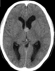
CT scan findings include a round mass with high attenuation at the anterior third ventricle. Rarely, colloid cysts may be isoattenuating or hypoattenuating relative to brain parenchyma. The typical high attenuation likely results from proteinaceous fluid. Hydrocephalus, which can be severe, results when the cyst obstructs the lateral ventricles at the foramen of Monro. This finding can be intermittent, as the cyst can act as a ball valve. Patients may present with intermittent positional headaches. A non-enhancing area of high attenuation in the typical location is almost diagnostic of a colloid cyst, but most clinicians confirm the diagnosis with MRI.