Choroid plexus papilloma
Choroid plexus papillomas are benign (WHO grade I), not very common neuroepithelial intraventricular tumors. These tumors are found both in children and adults, located inside the ventricles of the brain.
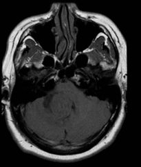
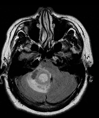
On imaging, these tumours are usually identified in the fourth ventricle in adults and in the lateral ventricles in the paediatric population. They commonly present as a solid vascular tumour with vivid frond-like enhancement pattern. In a quarter of cases, speckled calcifications are present.
Epidemiology
Overall these tumours account for approximately 1% of all brain tumours, 2-6% of all paediatric brain tumours and 0.5% of the adult brain tumours. They are, however, disproportionately represented in brain tumours in children under the age of 1. Approximately 85% of all choroid plexus papillomas occur in children under the age of 5 years.
Most cases present with hydrocephalus, believed to be due to a combination of overproduction of CSF and decreased arachnoid granulation resorption. These tumors have an association with Aicardi syndrome and Von Hippel-Lindau disease.
MRI features
Following contrast administration the frond-like morphology of the tumour can usually be
seen.
T1: typically isointense compared to adjacent brain; may be somewhat hypointense
T2: iso to hyperintense, small flow-voids may be seen within the tumor
T1 C+ (Gd): marked enhancement, tends to be homogeneous
Angiography: The tumors demonstrate an intense vascular blush on angiography. Sometimes enlarged choroidal arteries are seen feeding the tumor.