Ependymoma
An ependymoma is a tumor that arises from the ependyma, a tissue of the central nervous system. In pediatric cases the location is usually intracranial, while in adults it is spinal. The common location of intracranial ependymoma is the fourth ventricle. Rarely, an ependymoma may occur in the pelvic cavity.
Syringomyelia can be caused by an ependymoma. Ependymomas are also seen with neurofibromatosis type II.
Epidemiology
Ependymomas can occur at any age, though there is a peak in incidence around childhood (mean 6 years) and early adulthood (3rd decade). There is no gender predilection.
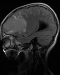
MRI features
Ependymomas are typically heterogeneous masses with areas of necrosis, calcification, cystic change and haemorrhage frequently seen. This results in a heterogeneous appearance on all modalities.
Intraparenchymal lesions (usually supratentorial) are usually large and variable in appearance, ranging from completely solid, enhancing masses to cysts with a mural nodule, or more heterogeneous masses.
T1: Solid portions of ependymoma typically are isointense to hypointense relative to white matter.
T2: Hyperintense compared to white matter. T2 weighted images are more reliable in differentiating tumor margins than non-contrast T1-weighted images (but less reliable than contrast enhanced T1).
SWI: Foci of blooming from haemorrhage or calcification may be seen.
T1 C+ (Gd): Enhancement is usually present but heterogeneous
DWI/ADC: Restricted diffusion may be seen in solid components, especially in anaplastic tumor. Diffusion should be interpreted with caution in masses with significant haemorrhage or calcification