Hemangioblastoma
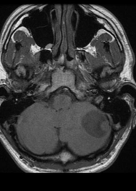
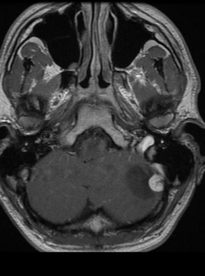
Hemangioblastomas are vascular tumors that can occur in different organs in the body, including liver, kidneys, pancreas, and the central nerve system. The tumors may be sporadic, or present in patients with von Hippel Lindau disease (in which they present at a younger age). Hemangioblastomas are low grade (WHO grade I) tumors that present on imaging as sharply demarcated homogeneous masses. Typical are a cystiform mass with non-enhancing walls, a mural nodule which brightly enhances with prominent flow voids. 95% of hemangioblastomas present in the posterior fossa.
Epidemiology
Hemangioblastomas typically occur in young to middle-aged adults with a peak incidence around 30-60 years of age. They are and, the most common primary posterior fossa mass in this demographic, though are uncommon in absolute numbers. Hemangioblastomas only account for 1-2.5% of all intracranial tumours and approximately 10% of all posterior fossa tumours. There is a slight male preponderance.
MRI features
T1: T1 imaging shows a hypointense to isointense mural nodule. The cystiform component shows CSF content.
T1 C+ (Gd): The mural nodule vividly enhances, the cyst wall does not enhance.
T2: T2 weighted imaging shows a hyperintense mural nodule with flow voids due to enlarged vessels, especially at the periphery of the cyst (60-70% of cases).
MR perfusion imaging: Being a highly vascular tumor, hemangioblastomas show high rCBV ratios on perfusion imaging