Metastases
Roughly 25-50% of all intracranial malignancies are estimated to be metastases in hospitalised patients. There is a great variation in imaging appearances, often presenting a diagnostic challenge.
Epidemiology
Five primary tumors account for 80% of brain metastases:
1. lung cancer
2. renal cell carcinoma
3. breast cancer
4. melanoma
5. gastrointestinal tract adenocarcinomas (the majority colorectal carcinoma)
MRI features
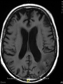
T1: Typically shows iso- to hypointense signal. Hemorrhagic lesions may have an intrinsic high signal. Non-hemorrhagic melanoma metastases may have a high signal do to the paramagnetic properties of melanin.
T1C+: The enhancement pattern can be uniform, punctate, or ring-enhancing, but it is usually intense. Delayed sequences may show additional lesions.
T2: T2 imaging is usually hyperintense, though hemorrhage may alter this.
FLAIR: FLAIR sequencing typically shows a hyperintense signal and hyperintense peri-tumoral oedema.
DWI/ADC: The oedema is out of proportion with tumour size and appears dark on DWI.
ADC demonstrates facilitated diffusion in oedema.


