Schwannoma
Intracranial schwannomas, or neurinomas, are fairly common benign tumors of the central nervous system. They make up 8% of all intracranial tumors, with the vast majority arising from the vestibular division of the vestibulocochlear cranial nerve. Schwannomas may occur anywhere in the body.
Epidemiology
Schwannomas are commonly found in middle-aged and elderly patients but may occur at any age. There is a female preponderance of about 1,5-2 : 1.
Almost all intracranial schwannomas arise from cranial nerves. Although any cranial nerve may be involved (with the exception of the olfactory nerve and optic nerve which lack sheaths made of Schwann cells), neurinomas of the vestibulocochlear nerve, trigeminal nerve, and facial nerve are most common.
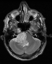
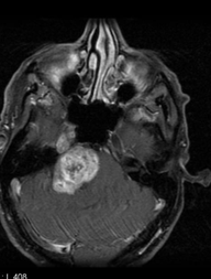
MRI features
MRI is the preferred method of diagnosis allowing for better anatomical localisation compared to (contrast) CT.
T1: The T1 weighted images show an isointense to hypointense lesion compared to brain. If present, the cystic areas show a low signal.
T1 C+ (Gd): Schwannomas tend to enhance prominently on contrast MRI, in most cases heterogeneously (70%).
T2: T2 imaging typically shows a somewhat hyperintense signal compared to brain tissue. The cystica areas if present, are hyperintense.
T2* (GE / SWI): Haemosiderin staining may be encountered, particularly in larger tumors.
DWI/ADC: Usually DWI/ADC shows a higher signal on both DWI and ADC (T2 shine through - not restricted diffusion)