Tumefactive multiple sclerosis
Tumefactive multiple sclerosis is a term used to describe patients with established multiple sclerosis who develop large aggressive demyelinating lesions, sometimes mimicing brain tumors or other space occupying intracranial lesions. They are similar/identical in appearance to tumefactive demyelinating lesions (TDL), also sometimes referred to as monofocal acute inflammatory demyelination (MAID), that are a distinct form of tumefactive lesions developing in patients without multiple sclerosis.
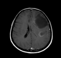
Tumefactive demyelinating lesions tend to be large but with relatively little mass effect or surrounding oedema. Centrally located dilated veins have also been observed within these lesions
MRI features
T1 C+ (Gd): About half of tumefactive demyelinating lesions demonstrate contrast enhancement. The enhancement pattern is usually in the form on an open ring and the incomplete portion of the ring is on the gray matter side of the lesion (open ring-enhancement)
Perfusion imaging helps differentiate tumefactive demyelinating lesions from the main differential considerations; high-grade gliomas and lymphomas. Mean relative cerebral blood volume within tumefactive demyelinating lesions have been found to be substantially less than in high-grade gliomas and lymphoma.
diffusion imaging (DWI): Is less specific in differentiating tumefactive demyelinating lesions from tumors, as necrotic neoplasms may display a similar increase in apparent diffusion coefficient (ADC). It may be used to differentiate between cerebral abscesses.